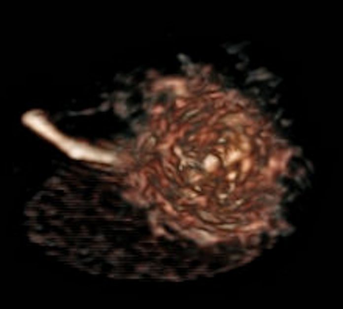
A red rose, picked from the garden at The Christie by a patient, has been chosen as the first item to be scanned by the hospital’s revolutionary new MR-linac radiotherapy machine.
The MR-guided linear accelerator (MR-linac) is currently one of only seven experimental systems in the world. It combines magnetic resonance (MR) scanning and tumour-busting radiotherapy to deliver magnetic resonance radiotherapy in one hi-tech package. It is expected to deliver some of the most precise radiotherapy available once fully operational.
The installation of the new £5.3m machine was completed earlier this year and since then researchers at The Christie have been testing the MR-linac.
The imaging part of the system is a vital component of the MR-linac and the successful scanning of the first item marks a major milestone in the development of magnetic resonance radiotherapy at The Christie.
Magnetic resonance radiotherapy is an advanced form of radiotherapy that will potentially enable The Christie to deliver more targeted treatment for patients with certain cancers.
The rose is England’s national flower and many patients at The Christie find peace and tranquillity amongst the roses in the hospital’s secluded garden. Prostate cancer patient Mike Thorpe (60) was invited to choose a rose from the hospital garden and then see it being scanned.
Mike was diagnosed with prostate cancer in 2014 and received radiotherapy at The Christie as part of his treatment. Prostate cancer is one of several types of cancer that will be treated with greater precision by the MR-linac once it is operational. Mike was involved in a clinical trial at The Christie and had his treatment in the research facility that the MR-linac is now replacing.
Mike commented, “It’s exciting to be involved in marking the development of the MR-linac at The Christie. For prostate cancer patients, the more accurate and precise the treatment, the better the long term survival and quality of life for us. The garden in the centre of The Christie is a tranquil place for patients and staff alike so it’s appropriate that the first item scanned by the MR-linac is a rose grown there.”
MR-linac can precisely locate tumours, tailor the shape of X-ray beams in real time, and lock-on to tumours during treatment, to deliver radiotherapy even when tumours are moving. It can also see if a tumour changes shape, location or size between treatment sessions.
Planning radiotherapy in “real time” avoids the need to rely on scans that might have been taken a week or two earlier.
The next major milestone for the MR-linac project will be the first scans of a human body which will be undertaken on a volunteer.
Dr Ananya Choudhury, clinical lead for MR-linac at The Christie, said: “The MR-linac is a flagship research project for The Christie using state-of-the-art experimental technology to deliver more precise, more personalised, more effective and kinder radiotherapy to our patients. It lets us see tumours very clearly and treat them at the same time with pinpoint accuracy.”


